2018-02-14
 350
350
Nervous tissue is composed of two main cell types: neurons and glial cells. Neurons transmit nerve messages. Glial cells are in direct contact with neurons and often surround them.
The neuron is the functional unit of the nervous system. Humans have about 100 billion neurons in their brain alone! While variable in size and shape, all neurons have three parts. Dendrites receive information from another cell and transmit the message to the cell body. The cell body contains the nucleus, mitochondria and other organelles typical of eukaryotic cells. The axon conducts messages away from the cell body.
Structure of a typical neuron. Three types of neurons occur. Sensory neurons typically have a long dendrite and short axon, and carry messages from sensory receptors to the central nervous system. Motor neurons have a long axon and short dendrites and transmit messages from the central nervous system to the muscles (or to glands). Interneurons are found only in the central nervous system where they connect neuron to neuron.
Synapses. The junction between a nerve cell and another cell is called a synapse. Messages travel within the neuron as an electrical action potential. The space between two cells is known as the synaptic cleft. Neurotransmitters are small molecules, some are even hormones. Neurotransmitters are either destroyed by specific enzymes in the synaptic cleft, or are reabsorbed by the cell. More than 30 organic molecules act as neurotransmitters.
Three basic functions are performed by nervous systems:
· Receive sensory input from internal and external environments
· Integrate the input
· Respond to stimuli
Sensory input. Receptors are parts of the nervous system that sense changes in the internal or external environments. Sensory input can be in many forms, including pressure, taste, sound, light, blood pH, or hormone levels that are converted to a signal and sent to the brain or spinal cord.
Integration and output. In the sensory centers of the brain or in the spinal cord, the barrage of input is integrated and a response is generated. The response, a motor output, is a signal transmitted to organs than can convert the signal into some form of action, such as movement, changes in heart rate, release of hormones, etc.
Endocrine systems. Some animals and humans beings have a second control system, the endocrine system. The nervous system coordinates rapid responses to external stimuli. The endocrine system controls slower, longer lasting responses to internal stimuli. Activity of both systems is integrated.
Divisions of the nervous system. The Central Nervous System (CNS) includes the brain and spinal cord. The Peripheral Nervous System (PNS) connects the CNS to other parts of the body, and is composed of nerves (bundles of neurons).
Not all animals have highly specialized nervous systems. Those with simple systems tend to be either small and very mobile or large and immobile. Large, mobile animals have highly developed nervous systems.
Two main components of the PNS are:
1. sensory (afferent) pathways that provide input from the body into the CNS.
2. motor (efferent) pathways that carry signals to muscles and glands (effectors).
The Somatic Nervous System (SNS) includes all nerves controlling the muscular system and external sensory receptors. External sense organs (including skin) are receptors. Muscle fibers and gland cells are effectors. The reflex arc is an automatic, involuntary reaction to a stimulus. The reaction to the stimulus is involuntary, with the CNS being informed but not consciously controlling the response. Examples of reflex arcs include balance, the blinking reflex, and the stretch reflex.
The Autonomic Nervous System is that part of PNS consisting of motor neurons that control internal organs. It has two subsystems. The autonomic system controls muscles in the heart, the smooth muscle in internal organs such as the intestine, bladder (мочевой пузырь), and uterus (матка). The Sympathetic Nervous System is involved in the fight or flight response. The Parasympathetic Nervous System is involved in relaxation. Each of these subsystems operates in the reverse of the other (antagonism). Both systems innervate the same organs and act in opposition to maintain homeostasis. For example: when you are scared the sympathetic system causes your heart to beat faster; the parasympathetic system reverses this effect.
Motor neurons in this system do not reach their targets directly (as do those in the somatic system) but rather connect to a secondary motor neuron which in turn innervates the target organ.
The Central Nervous System (CNS) is composed of the brain and spinal cord. The CNS is surrounded by bone-skull and vertebrae. Fluid and tissue also insulate the brain and spinal cord.
Areas of the brain
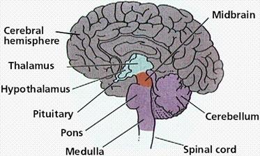 Fig.1
Fig.1
The brain is composed of three parts: the cerebrum (seat of consciousness), the cerebellum, and the medulla oblongata (these latter two are "part of the unconscious brain").
The medulla oblongata is closest to the spinal cord, and is involved with the regulation of heartbeat, breathing, vasoconstriction (blood pressure), and reflex centers for vomiting, coughing, sneezing, swallowing, and hiccuping. The hypothalamus regulates homeostasis. It has regulatory areas for thirst, hunger, body temperature, water balance, and blood pressure, and links the Nervous System to the Endocrine System. The midbrain and pons are also part of the unconscious brain. The thalamus serves as a central relay point for incoming nervous messages.
The cerebellum is the second largest part of the brain, after the cerebrum. It functions for muscle coordination and maintains normal muscle tone and posture. The cerebellum coordinates balance.
The conscious brain includes the cerebral hemispheres, which are separated by the corpus callosum. In reptiles, birds, and mammals, the cerebrum coordinates sensory data and motor functions. The cerebrum governs intelligence and reasoning, learning and memory. While the cause of memory is not yet definitely known, studies on slugs indicate learning is accompanied by a synapse decrease. Within the cell, learning involves change in gene regulation and increased ability to secrete transmitters.
The brain. During embryonic development, the brain first forms as a tube, the anterior end of which enlarges into three hollow swellings that form the brain, and the posterior of which develops into the spinal cord. Some parts of the brain have changed little during vertebrate evolutionary history.
The brain stem and midbrain. The brain stem is the smallest and from an evolutionary viewpoint, the oldest and most primitive part of the brain. The brain stem is continuous with the spinal cord, and is composed of the parts of the hindbrain and midbrain. The medulla oblongata and pons control heart rate, constriction of blood vessels, digestion and respiration.The midbrain consists of connections between the hindbrain and forebrain. Mammals use this part of the brain only for eye reflexes.
The cerebellum. The cerebellum is the third part of the hindbrain, but it is not considered part of the brain stem. Functions of the cerebellum include fine motor coordination and body movement, posture, and balance. This region of the brain is enlarged in birds and controls muscle action needed for flight.
The forebrain. The forebrain consists of the diencephalon and cerebrum. The thalamus and hypothalamus are the parts of the diencephalon. The thalamus acts as a switching center for nerve messages. The hypothalamus is a major homeostatic center having both nervous and endocrine functions.
The cerebrum, the largest part of the human brain, is divided into left and right hemispheres connected to each other by the corpus callosum. The hemispheres are covered by a thin layer of gray matter known as the cerebral cortex, the most recently evolved region of the vertebrate brain. Fish have no cerebral cortex, amphibians and reptiles have only rudiments of this area.
The cortex in each hemisphere of the cerebrum is between 1 and 4 mm thick. Folds divide the cortex into four lobes: occipital, temporal, parietal, and frontal. No region of the brain functions alone, although major functions of various parts of the lobes have been determined.
The major brain areas and lobes
 Fig.2
Fig.2
The occipital lobe (back of the head) receives and processes visual information. The temporal lobe receives auditory signals, processing language and the meaning of words. The parietal lobe is associated with the sensory cortex and processes information about touch, taste, pressure, pain, and heat and cold. The frontal lobe conducts three functions:
· motor activity and integration of muscle activity
· speech
· thought processes
Functional areas of the brain

|
Fig.3
Most people have their language and speech areas on the left hemisphere of their brain. Language comprehension is found in Wernicke’s area. Speaking ability is in Broca’s area. Damage to Broca’s area causes speech impairment but not impairment of language comprehension. Lesions in Wernicke’s area impairs ability to comprehend written and spoken words but not speech. The remaining parts of the cortex are associated with higher thought processes, planning, memory, personality and other human activities.