2020-08-05
 881
881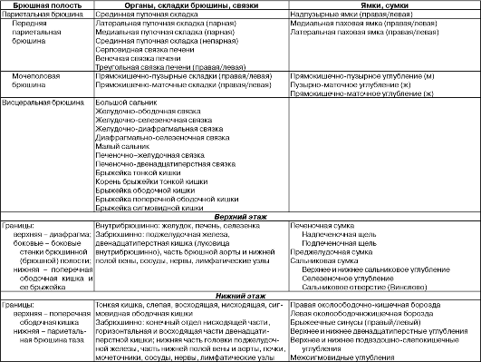
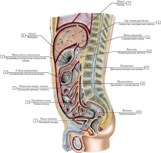 Рис. 185. Отношение внутренних органов к брюшине, срединный (сагиттальный) разрез туловища (схема):
1 - Urinary bladder; 2 - Small intestine; 3 - Greater omentum, cavity; 4 - Transverse colon; 5 - Transverse mesocolon; 6 - Stomach; 7 - Liver; 8 - Hepatogastric ligament; 9 - Omental bursa; Lesser sac; 10 - Pancreas; 11 - Duodenum; 12 - Mesentery; 13 - Rectum
 Рис. 186. Строение брюшины (схема)
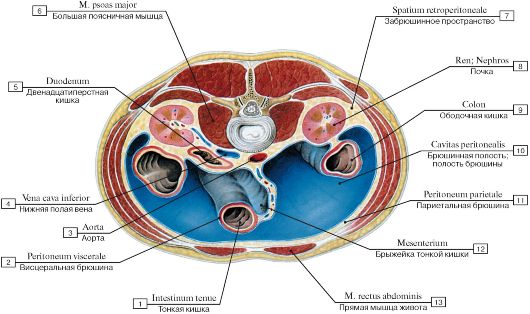 Рис. 187. Брюшная полость и расположенные в ней органы, горизонтальный (поперечный) распил туловища между телами II и III поясничных позвонков (схема):
1 - Small intestine; 2 - Visceral peritoneum; 3 - Aorta; 4 - Inferior vena cava; 5 - Duodenum; 6 - Psoas major; 7 - Retroperitoneal space; 8 - Kidney; 9 - Colon; 10 - Peritoneal cavity; 11 - Parietal peritoneum; 12 - Mesentery; 13 - Rectus abdominis
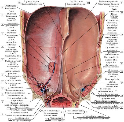 Рис. 188. Брюшина передней стенки живота, вид изнутри:
1 - Obturator internus; 2 - Supravesical fossa; 3 - Obturator artery and vein; 4 - Urinary bladder; 5 - Obturator nerve; 6 - Superior vesical artery; 7 - Ductus deferens; Vas deferens; 8 - Inguinal falx; Conjoint tendon; 9 - Spermatic cord; 10 - Interfoveolar ligament; 11 - Testicular artery and vein; 12 - Deep inguinal ring; 13 - Deep circumflex iliac artery and vein; 14 - Inferior epigastric artery and vein; 15 - Rectus abdominis; 16 - Arcuate line; 17 - Transverse fascia; 18 - Diaphragm; 19 - Round ligament of liver; 20 - Falciform ligament; 21 - Parietal peritoneum; 22 - Umbilical region; 23 - Transversus abdominis; Transverse abdominal; External and Internal oblique; 24 - Median umbilical ligament; 25 - Lateral umbilical fold; Epigastric fold; 26 - Iliopsoas; 27 - Lateral inguinal fossa; 28 - Femoral nerve; 29 - External iliac
artery and vein; 30 - Medial umbilical fold; 31 - Medial inguinal fossa
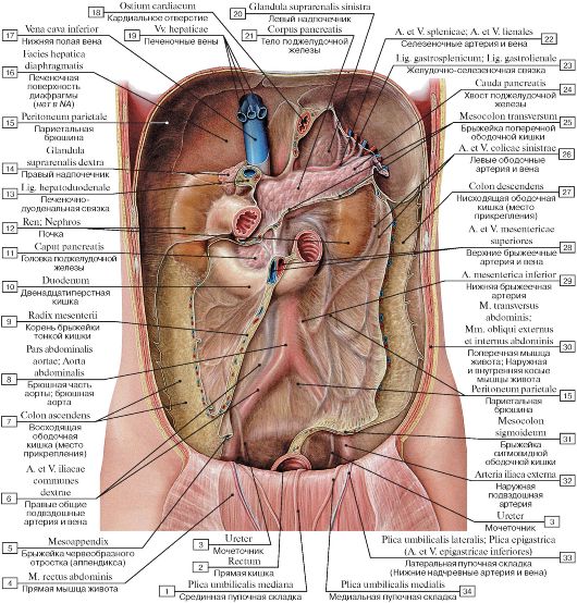 Рис. 189. Брюшина задней стенки живота, вид изнутри:
1 - Median umbilical fold; 2 - Rectum; 3 - Ureter; 4 - Rectus abdominis; 5 - Meso-appendix; 6 - Common iliac artery and vein; 7 - Ascending colon; 8 - Abdominal aorta; 9 - Root of mesentery; 10 - Duodenum; 11 - Head of pancreas; 12 - Kidney: 13 - Hepatoduodenal ligament; 14 - Right suprarenal gland; Right adrenal gland; 15 - Parietal peritoneum; 16 - Hepatic surface of diaphragm; 17 - Inferior vena cava; 18 - Cardial orifice; 19 - Hepatic veins; 20 - Left suprarenal gland; Left adrenal gland; 21 - Body of pancreas; 22 - Splenic artery and vein; 23 - Gastrosplenic ligament; 24 - Tail of pancreas; 25 - Transverse mesocolon; 26 - Left colic artery and vein; 27 - Descending colon; 28 - Superior mesenteric artery and vein; 29 - Inferior mesenteric artery; 30 - Transversus abdominis; Transverse abdominal; External and Internal oblique; 31 - Sigmoid mesocolon; 32 - External iliac artery; 33 - Lateral umbilical fold; Epigastric fold
(Inferior epigastric artery and vein); 34 - Medial umbilical fold
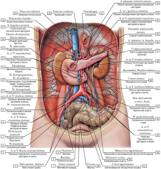 Рис. 190. Забрюшинное пространство:
1 - Urinary bladder; 2 - Rectum; 3 - Ovary; 4 - Uterine tube; 5 - Right external iliac artery and vein; 6 - Femoral nerve; 7 - Parietal peritoneum; 8 - Right common Шас artery and vein; 9 - Lateral cutaneous nerve of thigh; Lateral femoral cutaneous nerve; 10 - Right ovarian artery and vein; 11 - Ilio-inguinal nerve; 12 - Iliohypogastric nerve; Iliopubic nerve; 13 - Duodenum; 14 - Superior mesenteric artery and vein; 15 - Subcostal nerve; 16 - Bile duct; 17 - Hepatic artery proper; 18 - Hepatic portal vein; 19 - Inferior vena cava; 20 - Coeliac trunk; 21 - Oesophagus; 22 - Left inferior phrenic artery; 23 - Left gastric artery and vein; 24 - Superior suprarenal artery and vein; 25 - Left suprarenal gland; Left adrenal gland; 26 - Splenic artery; 27 - Body of pancreas; 28 - Left renal artery and vein; 29 - Left kidney; 30 - Left ureter; 31 - Left ovarian artery and vein; 32 - Abdominal aorta; 33 - Genitofemoral nerve; 34 - Inferior mesenteric artery and vein; 35 - Sigmoid mesocolon; 36 - Aortic bifurcation; 37 - Sigmoid colon; 38 - Uterus
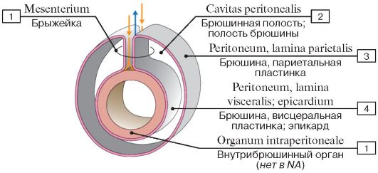 Рис. 191. Внутрибрюшинное расположение органов (схема):
1 - Mesentery; 2 - Peritoneal cavity; 3 - Peritoneum, parietal layer; 4 - Peritoneum, visceral layer; epicardium; 5 - Intraperitoneal organ
 Рис. 192. Большой сальник:
1 - Median umbilical fold; 2 - Arcuate line; 3 - Ileum; 4 - Caecum; 5 - Ascending colon; 6 - Free taenia; 7 - Greater omentum; 8 - Omental taenia; 9 - Transversus abdominis; Transverse abdominal; 10 - Fundus of gallbladder; 11 - Round ligament of liver; 12 - Right lobe of liver; 13 - Falciform ligament; 14 - Left lobe of liver; 15 - Body of stomach; 16 - Rectus abdominis; 17 - Pyloric part; 18 - Gastrocolic ligament; 19 - Stomach, greater curvature; 20 - Transverse colon; 21 - Transversus abdominis; Transverse abdominal; 22 - Internal oblique; 23 - External oblique; 24 - Parietal peritoneum; 25 - Lateral umbilical fold; Epigastric fold (Inferior epigastric artery and vein);
26 - Medial umbilical fold
 Рис. 193. Большой сальник (фотография нефиксированного натурального препарата):
1 - серповидная связка; 2 - вырезка круглой связки; 3 - сфинктер привратника; 4 = 19 - подсерозная основа; 5 - прямая мышца живота; 6 = 18 - париетальная брюшина; 7 = 16 - подкожная основа; гиподермис; 8 - левая доля печени, передняя часть диафрагмальной поверхности; 9 = 25 - печень, нижний край; 10 - тело желудка; 11 - большая кривизна желудка с правой желудочно-сальниковая артерией и ее желудочными ветвями; 12 - поперечная ободочная кишка, покрытая большим сальником; 13 - большой сальник; 14 - подвздошная кишка; 15 - паховая складка; паховая дуга; 17 - кожа; 20 - конечная часть подвздошной кишки; 21 - слепая кишка; 22 - свободная лента; 23 - гаустры ободочной кишки; 24 - желудочно-ободочная связка; 26 - дно
желчного пузыря; 27 - квадратная доля печени; 28 - круглая связка печени
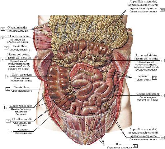 Рис. 194. Кишечник, большой сальник отвернут вверх:
1 - Caecum; 2 - Ileocaecal fold; 3 - Paracolic gutter; 4 - Free taenia; 5 - Ascending colon; 6 - Right colic flexure; Hepatic flexure; 7 - Free taenia; 8 - Transverse colon; 9 - Greater omentum; 10 - Omental appendices; Fatty appendices of colon; 11 - Left colic flexure;
Splenic flexure; 12 - Jejunum; 13 - Sigmoid colon; 14 - Ileum
 Рис. 195. Кишечник, большой сальник отвернут вверх (фотография нефиксированного натурального препарата):
1 - гаустры ободочной кишки; 2 - свободная лента; 3 - большой сальник; 4 = 15 - сальниковые отростки; 5 - брыжейка поперечной ободочной кишки; 6 - левый изгиб ободочной кишки; селезеночный изгиб ободочной кишки; 7 - сигмовидная ободочная кишка c лентами ободочной кишки и сальниковыми отростками; 8 - сигмовидная ободочная кишка; 9 = 13 - латеральная пупочная складка; 10 - срединная пупочная связка; 11 - тонкая кишка; 12 - пирамидальная мышца; 14 - слепая кишка;
16 - полулунная складка
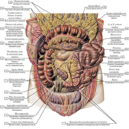 Рис. 196. Корень брыжейки тонкой кишки:
I - Superior and inferior ileocaecal recess; 2 - Ureter, abdominal part; 3 - Meso-appendix; 4 - Appendix; Vermiform appendix; 5 - Ileocaecal fold; 6 - Caecum; 7 - Paracolic gutter; 9 - Free taenia; 10 - Duodenum, inferior part; horizontal part; transverse part;
II - Right lobe of liver, inferior border; 12 - Right colic flexure; Hepatic flexure; 13 - Fundus of gallbladder; 14 - Haustra of colon; 15 - Greater omentum; 16 - Free taenia; 17 - Omental taenia; 18 - Middle colic artery and vein; 19 - Transverse mesocolon;
20 - Pancreas; 21 - Jejunum; 22 - Root of mesentery; 23 - Ileum, terminal ileum; 24 - Ileum
 Рис. 197. Корень брыжейки тонкой кишки (фотография нефиксированного натурального препарата):
1 - правая доля печени; 2 - корень брыжейки ободочной кишки (правая половина); 3 - корень брыжейки ободочной кишки (левая половина); 4 - большой сальник; 5 - поперечная ободочная кишка и сальниковые отростки; 6 - брыжейка поперечной ободочной кишки; 7 - двенадцатиперстно-тощекишечный изгиб; 8 - брыжейка тонкой кишки; 9 - верхнее илеоцекальное углубление; 10 - корень брыжейки тонкой кишки; 11 - изгиб подвздошной кишки; 12 - брыжейка тонкой кишки; 13 - брыжейка восходящей ободочной кишки; 14 - нижнее илеоцекальное углубление; 15 - брыжейка червеобразного отростка (аппендикса); 16 - червеобразный отросток; аппендикс; 17 - илеоцекальная складка; 18 - слепая кишка; 19 - свободная лента слепой кишки; 20 - сосудистая слепокишечная складка; 21 - правый изгиб ободочной кишки; печеночный изгиб ободочной кишки; 22 - париетальная брюшина; 23 - свободная лента поперечной ободочной кишки; 24 - сальниковые отростки: 25 - гаустры ободочной
кишки; 26 - диафрагма
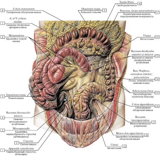 Рис. 198. Корень брыжейки тонкой кишки:
1 - Appendix; Vermiform appendix; 2 - Caecum; 3 - Meso-appendix; 4 - Inferior ileocaecal recess; 5 - Small intestine; 6 - Mesentery; 7 - Middle colic artery and vein; 8 - Transverse colon; 9 - Greater omentum; 10 - Free taenia; 11 - Pancreas, antero-inferior surface; 12 - Ureter; 13 - Superior and inferior duodenal fossa; 14 - Kidney, inferior pole; inferior extremity; 15 - Paracolic gutter; 16 - Sigmoid
colon; 18 - Intersigmoid recess; 18 - Sigmoid mesocolon
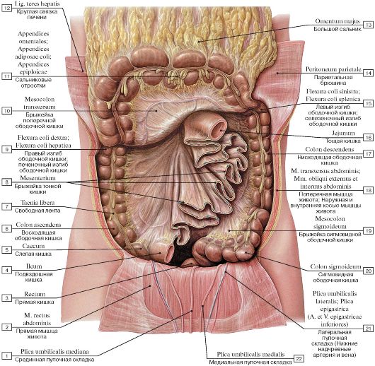 Рис. 199. Корень брыжейки тонкой кишки:
1 - Median umbilical fold; 2 - Rectus abdominis; 3 - Rectum; 4 - Ileum; 5 - Caecum; 6 - Ascending colon; 7 - Free taenia; 8 - Mesentery; 9 - Right colic flexure; Hepatic flexure; 10 - Transverse mesocolon; 11 - Omental appendices; Fatty appendices of colon; 12 - Round ligament of liver; 13 - Greater omentum; 14 - Parietal peritoneum; 15 - Left colic flexure; Splenic flexure; 16 - Jejunum; 17 - Descending colon; 18 - Transversus abdominis; Transverse abdominal; External oblique; Internal oblique; 19 - Sigmoid mesocolon; 20 - Sigmoid colon; 21 - Lateral umbilical fold; Epigastric fold (Inferior epigastric artery and vein); 22 - Medial umbilical fold
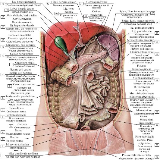 Рис. 200. Корень брыжейки тонкой кишки:
1 - Median umbilical fold; 2 - Rectus abdominis; 3 - Rectum; 4 - Ileum; 5 - Caecum; 6 - Ascending colon; 7 - Free taenia; 8 - Duodenum, inferior part; horizontal part; transverse part; 9 - Transverse colon; 10 - Right colic flexure; Hepatic flexure; 11 - Greater omentum; 12 - Stomach, pyloric part; 13 - Duodenum, superior part; 14 - Omental foramen; Epiploic foramen; 15 - Lesser omentum, hepatoduodenal ligament; 16 - Gallbladder; 17 - Right lobe of liver; 18 - Hepatogastric ligament; 19 - Left lobe of liver; 20 - Round ligament of liver; 21 - Body of pancreas; 22 - Cardial orifice; 23 - Spleen, gastric surface; 24 - Spleen, superior border; 25 - Gastrosplenic ligament; 26 - Transverse mesocolon; 27 - Left colic flexure; Splenic flexure; 28 - Duodenojejunal flexure; 29 - Descending colon; 30 - Transversus abdominis; Transverse abdominal; External and Internal oblique; 31 - Mesentery; 32 - Sigmoid mesocolon; 33 - Lateral umbilical fold;
Epigastric fold; 34 - Medial umbilical fold
 Рис. 201. Кишечник в брюшной полости, большой сальник отогнут кверху (фотография натурального препарата):
1 - Greater omentum; 2 - Left colic flexure; Splenic flexure; 3 - Transverse colon; 4 - Diaphragm; 5 - Liver; 6 - Descending colon; 7 - Jejunum; 8 - Middle colic artery; 9 - Right colic artery and vein; 10 - Superior mesenteric artery and vein; 11 - Intestinal vessels;
12 - Ileocolic artery and vein; 13 - Ascending colon; 14 - Caecum; 15 - Ileum
 Рис. 202. Органы брюшной полости, горизонтальный распил (схема):
1 - Descending colon; 2 - Paranephric fat; Pararenal fat body; 3 - Left colic flexure; Splenic flexure; 4 - Left kidney; 5 - Spleen; 6 - Pancreas; 7 - Omental bursa; Lesser sac; 8 - Splenic vein; 9 - Stomach, posterior wall; 10 - Stomach, anterior wall; 11 - Stomach, pyloric part; 12 - Greater omentum; 13 - Transverse colon; 14 - Duodenum; 15 - Superior mesenteric vein; 16 - Bile duct; 17 - Gallbladder; 18 - Superior mesenteric artery; 19 - Abdominal aorta; 20 - Inferior vena cava; 21 - Right lobe of liver; 22 - Intermediate lumbar nodes; 23 - Right suprarenal gland; Right adrenal gland; 24 - Right renal artery; 25 - Right kidney; 26 - Vertebra [LI]; 27 - Vertebral canal; Spinal cord; 28 - Lateral lumbar node
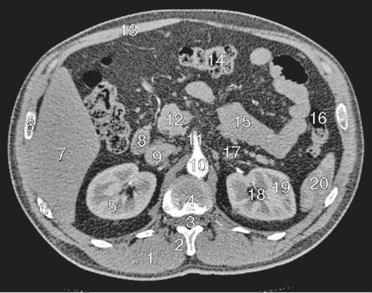 Рис. 203. Компьютерная томография брюшной полости:
I - длинная мышца спины; 2 - позвоночные мышцы; 3 - спинной мозг; 4 - грудной позвонок; 5 - почка (правая); 6 - ребра; 7 - правая доля печени; 8 - двенадцатиперстная кишка; 9 - нижняя полая вена; 10 - брюшная аорта;
II - верхняя брыжеечная артерия; 12-головка поджелудочной железы; 13 - прямая мышца живота; 14 - поперечная ободочная кишка; 15 - тонкий кишечник; 16 - нисходящая ободочная кишка; 17 - надпочечник (левый); 18 - верхние почечные чашечки; 19 - почка (левая); 20 - селезенка
(по С.К. Терновому)
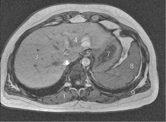 Рис. 204. Магнитно-резонансная томография брюшной полости, поперечный срез, Т1-взвешенное изображение:
1 - мышца-разгибатель спины; 2 - нижняя полая вена; 3 - правая доля печени; 4 - левая доля печени; 5 - пищевод; 6 - брюшная аорта; 7 - желудок; 8 - селезенка; 9 - широчайшая мышца спины (по С.К. Терновому)
 Рис. 205. Карманы и углубления брюшинной полости и таза мужчины, вид изнутри, спереди (схема)