2015-05-30
 381
381Оценка качества изображения должна выполняться одним из следующих методов:
1 Визуальный метод
Вылядит ли снимок качественным? Если снимок не выглядит хорошим, то, скорее всего, он не является качественным. Дефекты изображения и плотности, а также охват области исследования легко могут быть оценены визуально. Тем не менее, в случае оценки плотности, для оценки пограничных зон может потребоваться денситометр. Отпечатки пальцев, царапины на пленке или на экранах также часто становятся причинами, по которым снимок может быть забракован. В ряде случаев целесообразно обратиться к требованиям к определенным методам, но в любом случае здравый смысл поможет определить показания, которыми можно пренебречь.
2 Эталоны чувствительности (ЭЧ)/измерители проницаемости
Эталон чувствительности (ЭЧ), также называемый измерителем проницаемости, представляет собой вспомогательное устройство для оценки качества радиографического обследования при условиях измеримости малейших отклонений по толщине. На практике данное устройство добавляет плотности исследуемому образцу либо путем наложения проволочек, либо путем использования оптического клина, таким образом, чтобы малейшие колебания толщины могли быть определены. Затем данная величина выражается как процент от толщины образца и считается чувствительностью, выраженной в %. Если образец содержит трещину, следовательно, толщина исходного материала будет меньше в данной области, иначе говоря, объект будет иметь скачок плотности. Следует отметить, что когда достигается определенная чувствительность (или наблюдается изменение толщины), это еще не означает, что все неприемлемые дефекты определенного размера будут обнаружены. Проще всего это продемонстрировать на примере плоской трещины.
Везде, где это возможно ЭЧ следует размещать на стороне образца, обращенной к источнику, в области, которая с геометрической точки зрения является худшей. В качестве такой области может выступать самая плотная область образца или область, находящаяся недалеко от края диагностической пленки. Наиболее тонкая проволока должна быть расположена в наиболее удаленной от источника зоне.
| Рис. 1 | 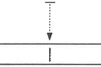
| Рис 2 | 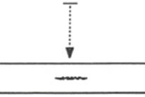
|
На рис. 1 показана такая трещина. В данном случае она расположена параллельно пучку излучения, и таким образом, создает резкий скачок плотности, что увеличивает возможность ее обнаружения.
На рис. 2 показана точно такая же трещина, но расположенная в плоскости перпендикулярной пучку. В данном случае это означает, что изменение плотности может быть недостаточным для обнаружения трещины. В обоих случаях можно предположить, что достижение максимальной чувствительности не означает, что все дефекты будут обнаружены.
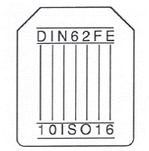
Проволочные ЭЧ
Данный тип эталона наиболее часто применяется вследствие малого веса и значительного диапазона толщин.
 Данные эталоны могут размещаться поперек сварного шва.
Они представляют собой 7 проволочек различной толщины, заключенных в три пластиковых оболочки. Каждая проволочка имеет собственное обозначение (номер от 1 до 16 или 18), которое соответствует определенной толщине проволочки. Оболочка затем обозначается в соответствии с номерами самой тонкой и самой толстой проволочки, которые она содержит (например, 10-16).
| DIN 62 | BS3971 1 Visually Does the image look alright? If the image does not appear to be so then the chances are that it is not. Density, image fall oft and inspection area coverage can all be quickly assessed visually. Although for density, borderline cases will need assessing using a densitometer. Blemishes, film scratches, screen scratches (see section entitled 'Artefacts') are often cause for rejecting the radiograph. It is advisable to refer to the specific process specifications in these cases but even so common sense is usually one of the best reasons for rejecting such indications. 2 Image quality indicators (IQI)/penetrameter An image quality indicator (IQI), sometimes called a penetrameter is a device which helps us to assess the radiographic quality in terms of the smallest thickness change detectable. In practice the device adds thickness to the test piece, either by overlaying wire or a stepwedge, so that the thinnest discernible change in thickness can be determined. This is then termed as a percentage of the testpiece thickness and given as the % sensitivity, tf an item contains a flaw it follows that the thickness of the parent material will be thinner in that area, in other words there is a change in thickness of the material. Care must be taken here to note that just because a sensitivity (or thickness change) has been measured with an IQI it does not always follow that all rejectable flaws down to that size will be detected. This is most easily shown with a planar type flaw. Wherever possible IQIs are placed on the source side of the specimen at the area considered to be the worst point geometrically. This could be on the thickest area of the specimen or at the edge of the diagnostic film length. The smallest wire should be placed at the furthest point away from the source. Figure 1 shows such a flaw. In this case the large dimension of the flaw is in line with the radiation beam and so creates a large thickness change in that area, it is likely that this will be detected. 1 Visually Does the image look alright? If the image does not appear to be so then the chances are that it is not. Density, image fall oft and inspection area coverage can all be quickly assessed visually. Although for density, borderline cases will need assessing using a densitometer. Blemishes, film scratches, screen scratches (see section entitled 'Artefacts') are often cause for rejecting the radiograph. It is advisable to refer to the specific process specifications in these cases but even so common sense is usually one of the best reasons for rejecting such indications. 2 Image quality indicators (IQI)/penetrameter An image quality indicator (IQI), sometimes called a penetrameter is a device which helps us to assess the radiographic quality in terms of the smallest thickness change detectable. In practice the device adds thickness to the test piece, either by overlaying wire or a stepwedge, so that the thinnest discernible change in thickness can be determined. This is then termed as a percentage of the testpiece thickness and given as the % sensitivity, tf an item contains a flaw it follows that the thickness of the parent material will be thinner in that area, in other words there is a change in thickness of the material. Care must be taken here to note that just because a sensitivity (or thickness change) has been measured with an IQI it does not always follow that all rejectable flaws down to that size will be detected. This is most easily shown with a planar type flaw. Wherever possible IQIs are placed on the source side of the specimen at the area considered to be the worst point geometrically. This could be on the thickest area of the specimen or at the edge of the diagnostic film length. The smallest wire should be placed at the furthest point away from the source. Figure 1 shows such a flaw. In this case the large dimension of the flaw is in line with the radiation beam and so creates a large thickness change in that area, it is likely that this will be detected. 1 Visually Does the image look alright? If the image does not appear to be so then the chances are that it is not. Density, image fall oft and inspection area coverage can all be quickly assessed visually. Although for density, borderline cases will need assessing using a densitometer. Blemishes, film scratches, screen scratches (see section entitled 'Artefacts') are often cause for rejecting the radiograph. It is advisable to refer to the specific process specifications in these cases but even so common sense is usually one of the best reasons for rejecting such indications. 2 Image quality indicators (IQI)/penetrameter An image quality indicator (IQI), sometimes called a penetrameter is a device which helps us to assess the radiographic quality in terms of the smallest thickness change detectable. In practice the device adds thickness to the test piece, either by overlaying wire or a stepwedge, so that the thinnest discernible change in thickness can be determined. This is then termed as a percentage of the testpiece thickness and given as the % sensitivity, tf an item contains a flaw it follows that the thickness of the parent material will be thinner in that area, in other words there is a change in thickness of the material. Care must be taken here to note that just because a sensitivity (or thickness change) has been measured with an IQI it does not always follow that all rejectable flaws down to that size will be detected. This is most easily shown with a planar type flaw. Wherever possible IQIs are placed on the source side of the specimen at the area considered to be the worst point geometrically. This could be on the thickest area of the specimen or at the edge of the diagnostic film length. The smallest wire should be placed at the furthest point away from the source. Figure 1 shows such a flaw. In this case the large dimension of the flaw is in line with the radiation beam and so creates a large thickness change in that area, it is likely that this will be detected. 1 Visually Does the image look alright? If the image does not appear to be so then the chances are that it is not. Density, image fall oft and inspection area coverage can all be quickly assessed visually. Although for density, borderline cases will need assessing using a densitometer. Blemishes, film scratches, screen scratches (see section entitled 'Artefacts') are often cause for rejecting the radiograph. It is advisable to refer to the specific process specifications in these cases but even so common sense is usually one of the best reasons for rejecting such indications. 2 Image quality indicators (IQI)/penetrameter An image quality indicator (IQI), sometimes called a penetrameter is a device which helps us to assess the radiographic quality in terms of the smallest thickness change detectable. In practice the device adds thickness to the test piece, either by overlaying wire or a stepwedge, so that the thinnest discernible change in thickness can be determined. This is then termed as a percentage of the testpiece thickness and given as the % sensitivity, tf an item contains a flaw it follows that the thickness of the parent material will be thinner in that area, in other words there is a change in thickness of the material. Care must be taken here to note that just because a sensitivity (or thickness change) has been measured with an IQI it does not always follow that all rejectable flaws down to that size will be detected. This is most easily shown with a planar type flaw. Wherever possible IQIs are placed on the source side of the specimen at the area considered to be the worst point geometrically. This could be on the thickest area of the specimen or at the edge of the diagnostic film length. The smallest wire should be placed at the furthest point away from the source. Figure 1 shows such a flaw. In this case the large dimension of the flaw is in line with the radiation beam and so creates a large thickness change in that area, it is likely that this will be detected. 1 Visually Does the image look alright? If the image does not appear to be so then the chances are that it is not. Density, image fall oft and inspection area coverage can all be quickly assessed visually. Although for density, borderline cases will need assessing using a densitometer. Blemishes, film scratches, screen scratches (see section entitled 'Artefacts') are often cause for rejecting the radiograph. It is advisable to refer to the specific process specifications in these cases but even so common sense is usually one of the best reasons for rejecting such indications. 2 Image quality indicators (IQI)/penetrameter An image quality indicator (IQI), sometimes called a penetrameter is a device which helps us to assess the radiographic quality in terms of the smallest thickness change detectable. In practice the device adds thickness to the test piece, either by overlaying wire or a stepwedge, so that the thinnest discernible change in thickness can be determined. This is then termed as a percentage of the testpiece thickness and given as the % sensitivity, tf an item contains a flaw it follows that the thickness of the parent material will be thinner in that area, in other words there is a change in thickness of the material. Care must be taken here to note that just because a sensitivity (or thickness change) has been measured with an IQI it does not always follow that all rejectable flaws down to that size will be detected. This is most easily shown with a planar type flaw. Wherever possible IQIs are placed on the source side of the specimen at the area considered to be the worst point geometrically. This could be on the thickest area of the specimen or at the edge of the diagnostic film length. The smallest wire should be placed at the furthest point away from the source. Figure 1 shows such a flaw. In this case the large dimension of the flaw is in line with the radiation beam and so creates a large thickness change in that area, it is likely that this will be detected. 1 Visually Does the image look alright? If the image does not appear to be so then the chances are that it is not. Density, image fall oft and inspection area coverage can all be quickly assessed visually. Although for density, borderline cases will need assessing using a densitometer. Blemishes, film scratches, screen scratches (see section entitled 'Artefacts') are often cause for rejecting the radiograph. It is advisable to refer to the specific process specifications in these cases but even so common sense is usually one of the best reasons for rejecting such indications. 2 Image quality indicators (IQI)/penetrameter An image quality indicator (IQI), sometimes called a penetrameter is a device which helps us to assess the radiographic quality in terms of the smallest thickness change detectable. In practice the device adds thickness to the test piece, either by overlaying wire or a stepwedge, so that the thinnest discernible change in thickness can be determined. This is then termed as a percentage of the testpiece thickness and given as the % sensitivity, tf an item contains a flaw it follows that the thickness of the parent material will be thinner in that area, in other words there is a change in thickness of the material. Care must be taken here to note that just because a sensitivity (or thickness change) has been measured with an IQI it does not always follow that all rejectable flaws down to that size will be detected. This is most easily shown with a planar type flaw. Wherever possible IQIs are placed on the source side of the specimen at the area considered to be the worst point geometrically. This could be on the thickest area of the specimen or at the edge of the diagnostic film length. The smallest wire should be placed at the furthest point away from the source. Figure 1 shows such a flaw. In this case the large dimension of the flaw is in line with the radiation beam and so creates a large thickness change in that area, it is likely that this will be detected. 1 Visually Does the image look alright? If the image does not appear to be so then the chances are that it is not. Density, image fall oft and inspection area coverage can all be quickly assessed visually. Although for density, borderline cases will need assessing using a densitometer. Blemishes, film scratches, screen scratches (see section entitled 'Artefacts') are often cause for rejecting the radiograph. It is advisable to refer to the specific process specifications in these cases but even so common sense is usually one of the best reasons for rejecting such indications. 2 Image quality indicators (IQI)/penetrameter An image quality indicator (IQI), sometimes called a penetrameter is a device which helps us to assess the radiographic quality in terms of the smallest thickness change detectable. In practice the device adds thickness to the test piece, either by overlaying wire or a stepwedge, so that the thinnest discernible change in thickness can be determined. This is then termed as a percentage of the testpiece thickness and given as the % sensitivity, tf an item contains a flaw it follows that the thickness of the parent material will be thinner in that area, in other words there is a change in thickness of the material. Care must be taken here to note that just because a sensitivity (or thickness change) has been measured with an IQI it does not always follow that all rejectable flaws down to that size will be detected. This is most easily shown with a planar type flaw. Wherever possible IQIs are placed on the source side of the specimen at the area considered to be the worst point geometrically. This could be on the thickest area of the specimen or at the edge of the diagnostic film length. The smallest wire should be placed at the furthest point away from the source. Figure 1 shows such a flaw. In this case the large dimension of the flaw is in line with the radiation beam and so creates a large thickness change in that area, it is likely that this will be detected. 1 Visually Does the image look alright? If the image does not appear to be so then the chances are that it is not. Density, image fall oft and inspection area coverage can all be quickly assessed visually. Although for density, borderline cases will need assessing using a densitometer. Blemishes, film scratches, screen scratches (see section entitled 'Artefacts') are often cause for rejecting the radiograph. It is advisable to refer to the specific process specifications in these cases but even so common sense is usually one of the best reasons for rejecting such indications. 2 Image quality indicators (IQI)/penetrameter An image quality indicator (IQI), sometimes called a penetrameter is a device which helps us to assess the radiographic quality in terms of the smallest thickness change detectable. In practice the device adds thickness to the test piece, either by overlaying wire or a stepwedge, so that the thinnest discernible change in thickness can be determined. This is then termed as a percentage of the testpiece thickness and given as the % sensitivity, tf an item contains a flaw it follows that the thickness of the parent material will be thinner in that area, in other words there is a change in thickness of the material. Care must be taken here to note that just because a sensitivity (or thickness change) has been measured with an IQI it does not always follow that all rejectable flaws down to that size will be detected. This is most easily shown with a planar type flaw. Wherever possible IQIs are placed on the source side of the specimen at the area considered to be the worst point geometrically. This could be on the thickest area of the specimen or at the edge of the diagnostic film length. The smallest wire should be placed at the furthest point away from the source. Figure 1 shows such a flaw. In this case the large dimension of the flaw is in line with the radiation beam and so creates a large thickness change in that area, it is likely that this will be detected. 1 Visually Does the image look alright? If the image does not appear to be so then the chances are that it is not. Density, image fall oft and inspection area coverage can all be quickly assessed visually. Although for density, borderline cases will need assessing using a densitometer. Blemishes, film scratches, screen scratches (see section entitled 'Artefacts') are often cause for rejecting the radiograph. It is advisable to refer to the specific process specifications in these cases but even so common sense is usually one of the best reasons for rejecting such indications. 2 Image quality indicators (IQI)/penetrameter An image quality indicator (IQI), sometimes called a penetrameter is a device which helps us to assess the radiographic quality in terms of the smallest thickness change detectable. In practice the device adds thickness to the test piece, either by overlaying wire or a stepwedge, so that the thinnest discernible change in thickness can be determined. This is then termed as a percentage of the testpiece thickness and given as the % sensitivity, tf an item contains a flaw it follows that the thickness of the parent material will be thinner in that area, in other words there is a change in thickness of the material. Care must be taken here to note that just because a sensitivity (or thickness change) has been measured with an IQI it does not always follow that all rejectable flaws down to that size will be detected. This is most easily shown with a planar type flaw. Wherever possible IQIs are placed on the source side of the specimen at the area considered to be the worst point geometrically. This could be on the thickest area of the specimen or at the edge of the diagnostic film length. The smallest wire should be placed at the furthest point away from the source. Figure 1 shows such a flaw. In this case the large dimension of the flaw is in line with the radiation beam and so creates a large thickness change in that area, it is likely that this will be detected. | Диаметр проволоки (мм) | |||||
| 1 -7 | 6-12 | 10-16 | 1-7 | 4-10 | 9-15 | 15-21 | |
| *1 | 0.032 | ||||||
| '2 | 0.040 | ||||||
| '3 | 0.050 | ||||||
| 0.063 | |||||||
| 0.080 | |||||||
| 0.1 | |||||||
| 0.125 | |||||||
| 0.160 | |||||||
| 0.2 | |||||||
| 0.25 | |||||||
| 0.32 | |||||||
| 0.4 | |||||||
| 0.5 | |||||||
| 0.63 | |||||||
| OS | |||||||
| 1.0 | |||||||
| 1.25 | |||||||
| 4 | 1.6 | ||||||
| 2.0 | |||||||
| 2.5 | |||||||
| 3.2 |
* Данные проволочки изготавливаются только под заказ.
Толщина самой тонкой проволочки, видимой на радиографическом снимке, может быть определена. Чувствительность, выраженную в процентах можно определить с учетом толщины материала по приведенной ниже формуле:

Например, в случае, если проволочка с обозначением 10 видна на участке объекта толщиной 50мм при использовании ЭЧ 6-12Б DIN 62, то в соответствии с указанной выше формулой чувствительность данного эталона определяется следующим образом:
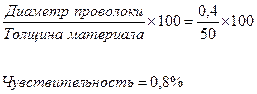
Некоторые эталоны позволяют достичь чувствительности до 2% при толщине материала до 50 мм и 1% при толщине материала свыше 50мм.
Следует обратить внимание: чем ниже процент чувствительности, тем она лучше.